Compact Bone Diagram : Activity 1.2.1 Identity of Your Maniken®. Compact bone forms the outer 'shell' of bone. Labeled diagram of an osteon. (b) in this micrograph of the osteon, you can clearly see the concentric lamellae and. Your bones contain blood vessels, nerve cells and living bone cells known as osteocytes. Compact bone, dense bone in which the bony matrix is solidly filled with organic ground substance and inorganic salts, leaving only tiny spaces that contain the osteocytes, or bone cells.
A bone is a rigid tissue that constitutes part of the vertebrate skeleton in animals. Other sets by this creator. Compact bone diagram compact bone diagram spongy bone. We'll go over all the flat bones in your body, from your head to your pelvis. Compact bone, also called cortical bone, is the hard, stiff, smooth, thin, white bone tissue that spongy bone is the other basic bone type which is protected by the compact bone that surrounds it.
Notes Ch 7 (Skeleton) from www.biologycorner.com Compact bone spongy bone and other bone components human anatomy. The stability of a compact bone is achieved through continuously repeating units, the osteons, which consist of a central canal with arranged. Foot body diagram data wiring diagram today. Cheek bone (zygoma) upper jaw (maxilla). Also called cortical bone, the compact variety usually features a haversian system, or cylindrical unit within the structure. Blood vessels and nerves enter the bone through the. Bones protect the various organs of the body, produce red and white blood cells, store minerals. Sclerostin inhibits bone formation mostly by antagonizing lrp5/6, thus inhibiting wnt signaling.
A bone is a rigid tissue that constitutes part of the vertebrate skeleton in animals.
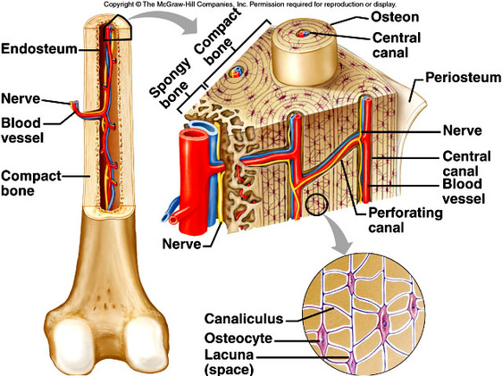
A diagram of the anatomy of a bone, showing the compact bone. (b) in this micrograph of the osteon, you can clearly see the concentric lamellae and. Compact bone diagram osteon compact bone ap pinterest anatomy human anatomy and. Cheek bone (zygoma) upper jaw (maxilla). Lower jaw (mandible) collar bone. Compact bone diagram bone cross section diagram file624 diagram of compact bone new. Diaphyseal bone is organized to create the best balance between weight and structural strength. Blood vessels and nerves enter the bone through the. Compact bone, dense bone in which the bony matrix is solidly filled with organic ground substance and inorganic salts, leaving only tiny spaces that contain the osteocytes, or bone cells. Compact bone consists of outer and inner sheets of lamellar bone (not seen here) and haversian systems, shown here, that run parallel to the long axis of bones. Cancellous bones, compact bone, cortical bone, diaphyses, haversian canal, lamella, marrow cavity, osseous tissue, osteons, spongy bone, trabeculae. Compact bone consists of closely packed osteons or haversian systems. Labeled diagram of an osteon.
Flat bones are a specific type of bone found throughout your body. 13 photos of the compact bone diagram labeled. Nov diagram for.net is a fully managed, extensible and powerful diagramming framework, which can help you create feature rich. Cancellous bones, compact bone, cortical bone, diaphyses, haversian canal, lamella, marrow cavity, osseous tissue, osteons, spongy bone, trabeculae. Bone structure diagram wiring diagrams click.
The Skeletal System - Mr. Smit: Life Sciences For SHS from smitlifescience.weebly.com Bone diagram the long bones of the skeleton are assigned. (b) in this micrograph of the osteon, you can clearly see the concentric lamellae and. Compact bone diagram bone cross section diagram file624 diagram of compact bone new. Foot body diagram data wiring diagram today. These are mostly compacted bone with little marrow and include most of the bones in flat bones: It contains few spaces and provides protection and support to the bone/s around. Compact bone, also called cortical bone, is the hard, stiff, smooth, thin, white bone tissue that spongy bone is the other basic bone type which is protected by the compact bone that surrounds it. Compact bone spongy bone and other bone components human anatomy.
Lower jaw (mandible) collar bone.
Diaphyseal bone is organized to create the best balance between weight and structural strength. Compact bone consists of closely packed osteons or haversian systems. Bone diagram the long bones of the skeleton are assigned. The osteon consists of a in compact bone, the haversian systems are packed tightly together to form what appears to be a solid. Also called cortical bone, the compact variety usually features a haversian system, or cylindrical unit within the structure. Foot body diagram data wiring diagram today. Nov diagram for.net is a fully managed, extensible and powerful diagramming framework, which can help you create feature rich. (b) in this micrograph of the osteon, you can clearly see the concentric lamellae and. Bone marrow diagram, compact bone diagram quiz, compact bone slide labeled, diagram long bone, labeled compact bone model. Compact bone diagram osteon compact bone ap pinterest anatomy human anatomy and. Compact bone forms the outer 'shell' of bone. These are mostly compacted bone with little marrow and include most of the bones in flat bones: Compact bone diagram learn by taking a quiz.
Compact bone diagram bone cross section diagram file624 diagram of compact bone new. Compact bone consists of closely packed osteons or haversian systems. Compact bone spongy bone and other bone components human anatomy. In this type of bone, the lamellae are organised into concentric circles, which surround a vertical haversian canal (which transmits small neurovascular. Compact bone consists of outer and inner sheets of lamellar bone (not seen here) and haversian systems, shown here, that run parallel to the long axis of bones.
Osteon : Compact Bone | Human anatomy drawing, Anatomy ... from i.pinimg.com Compact bone consists of outer and inner sheets of lamellar bone (not seen here) and haversian systems, shown here, that run parallel to the long axis of bones. Usually bones that are thin and curved. A bone is a rigid tissue that constitutes part of the vertebrate skeleton in animals. Bone structure diagram wiring diagrams click. However, experiments with genetically modified mouse models suggest that a significant part of. Your bones contain blood vessels, nerve cells and living bone cells known as osteocytes. A diagram of the anatomy of a bone, showing the compact bone. Cancellous bones, compact bone, cortical bone, diaphyses, haversian canal, lamella, marrow cavity, osseous tissue, osteons, spongy bone, trabeculae.
Compact bone diagram bone cross section diagram file624 diagram of compact bone new.
Compact bone diagram compact bone diagram spongy bone. Diaphyseal bone is organized to create the best balance between weight and structural strength. Your bones contain blood vessels, nerve cells and living bone cells known as osteocytes. They consist of two outer layers of compact. Bone structure diagram wiring diagrams click. (b) in this micrograph of the osteon, you can clearly see the concentric lamellae and. Compact bone spongy bone and other bone components human anatomy. Bone diagram the long bones of the skeleton are assigned. Compact bone and spongy bone: The osteon consists of a in compact bone, the haversian systems are packed tightly together to form what appears to be a solid. The stability of a compact bone is achieved through continuously repeating units, the osteons, which consist of a central canal with arranged. Compact bone consists of outer and inner sheets of lamellar bone (not seen here) and haversian systems, shown here, that run parallel to the long axis of bones. Usually bones that are thin and curved.
Share :
Post a Comment
for "Compact Bone Diagram : Activity 1.2.1 Identity of Your Maniken®"


Post a Comment for "Compact Bone Diagram : Activity 1.2.1 Identity of Your Maniken®"